Optical
Imaging Core
 Optical Imaging Core
The Optical Imaging Core provides imaging services for Van Andel Institute and collaborating institutions. Its goal is to serve the VAI community with state-of-the-art imaging resources to address a broad variety of research questions — from gene expression to cell motility — as well as offering the expertise to train researchers to use these resources effectively.
How to request services
VAI’s Core Technologies and Services manages service requests through CrossLab Solutions. Each Core maintains an CrossLab page that provides descriptions of the Core as well as options to schedule equipment and request services. Pricing also is available in CrossLab (See Schedule Equipment and/or Request Services). Current CrossLab users can login here.
VAI staff that need a login (or further CrossLab support) should contact Lori Moon. MSU and external users that need a CrossLab account/login should go here for account creation. After logging in, external users should click “list all cores” under the Core Facilities section in the left menu and scroll down to “Cores at Van Andel Institute.”
Please request all services through CrossLab. Contact Dr. Corinne Esquibel with any questions.
Request Genomics, Infinium, and Single Cell Sequencing Services
Spatial Collaborative Services combines expertise and technology from VAI’s Genomics Core and Optical Imaging Core to offer investigators a seamless pipeline for spatial imaging. For questions, please email [email protected].
Our Impact
We’re raising thousands to save millions.
We’re turning hope into action for the millions of people around the world affected by diseases like cancer and Parkinson’s. Find out how you can help us make a difference.
- 141 peer-reviewed papers published in 2025, 74 of which were in high-impact journals
- 15 VAI-SU2C Epigenetics Dream Team clinical trials launched to date
- 10 clinical trials and related projects supported by VAI through the International Linked Clinical Trials Program
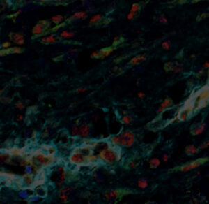
The Optical Imaging Core provides state-of-the-art microscopy services for biological specimens. Core capabilities are used to explore a wide array of experimental questions, and includes (but not limited to):
-
- Fluorescent imaging
- Brightfield imaging
- Optical sectioning
- 3-D rendering
- Live-cell imaging
- Spectral unmixing
- Fluorescence recovery after photobleaching (FRAP)
- Photoactivation and photoconversion
- Colocalization
- Second harmonic generation
- Slide digitization
- In vivo screening
- Multi-photon Imaging
- Total Internal Reflection Fluorescence (TIRF)
- Structured Illumination Microscopy
- Automated CyCIF Staining and Imaging
Training
Training in equipment operation and imaging data analysis is available. Contact [email protected] to schedule training.
How to Access Services
New users should complete training before signing up or accessing the confocal suite.
Certified internal users should login to CrossLab and schedule service times through the system. New users who need login information should contact Lori Moon.
Equipment
- Axio Observer 7 inverted microscope body
- Definite Focus 2
- Automated scanning stage
- High-speed Z-piezo frame
- Stage-surround incubation provides temperature, humidity and gas controls for live-cell imaging
- 5x air, 10x air, 20x air, 40x water, 63x oil and 100x oil objectives
- Airyscan detector
- Airyscan superresolution mode
- Airyscan Fast mode
- 405, 458, 488, 514, 561 and 633 nm laser lines for confocal imaging
- Confocal imaging detectors:
- Two liquid-cooled MA-PMT
- One high-sensitivity 32-channel GaAsP array
- Coherent Discovery dual-output Ti-Sapphire laser for multiphoton imaging
- Tunable output: 660–1320 nm
- Fixed output: 1040 nm
- Multiphoton imaging detectors (non-descanned):
- Two GaAsP
- Two multi-alkali PMT
- Transmitted light detector
- Zeiss Zen software
- Offline processing computer
- Leica DMi8 inverted microscope body
- Perfect Focus system
- Drift stabilization
- High speed Z-piezo
- 405, 488, 561, 640, and 730 nm laser lines
- 10x air, 20x air, 25x water, 40x oil, 63x oil and 100x oil objectives
- 40 um and 25 um pinholes
- Two Zyla 4.2MP sCMOS cameras
- Oko-Lab Stage-top incubator provides temperature, humidity, and gas controls for live-cell imaging
- CoolLED epifluorescence illumination
- CoolLED transmitted illumination
- Inserts for multiple sample types
- 35 mm dishes
- Slides
- Multi-well slides
- Multi-well plates
- Oko-Lab PF35 perfusion chamber
- Fusion acquisition software
- Imaris Core visualization software
- “Confocal-like” structured illumination, fully motorized, inverted widefield microscope with AI sample finder.
- Instrument is capable of several types of imaging:
- Brightfield
- Fluorescence
- DIC
- Polarization
- Stage inserts can accommodate:
- Slides
- Multi-well slides
- 35mm dishes
- Multi-well plates
- Fully automated slide scanning, 100 slide capacity
- Fluorescence
- Brightfield
- Polarization
- Z-stacking
- Colibri 7 VIS-LED light source
- 5x air, 10x air, 20x air, and 40x air objectives
- Zeiss Zen software
- Fully automated slide scanning (100 slide capacity):
- Fluorescence
- Brightfield
- Z-stacking
- Colibri 7 VIS-LED light source
- 5x air, 10x air, 20x air and 40x air objectives
- Fast filter wheel
- Zeiss Zen software
- Fully automated system for live-cell imaging
- Phase contrast gradient
- Fluorescence
- Time-course
- Z-stacking
- Tiling
- Full CO2 and heating control
- Definite focus
- Inserts for multiple sample types
- 35mm dishes
- 60mm dishes
- Slides
- Multi-well slides
- Multi-well plates
- Pecon POC-R2 perfusion chamber
- Autodetection of sample vessel
- Autocorrection of objectives to accommodate sample vessel thickness (plastic or glass)
- 5x air, 20x air, 20x water and 50x water objectives
- 5x, 1x and 2x magnification
- Autoimmersion for water objectives
- Zeiss Zen software
- Commercial software:
- Imaris
- SVI Huygens Deconvolution
- Zen Desktop
- Nikon Elements
- InForm
- Aperio ImageScope
- Matlab
- Freeware:
- FIJI/ImageJ
- CellProfiler
- CellProfiler Analyst
- QuPath
- CellPose
- Ilastik
- Anaconda Python Environment Manager
- Behavioral Observation Research Interactive Software (BORIS)
Courses and Workshops
- BioImaging North America
- BioImaging offers an extensive library of training and education resources.
- iBiology Microscopy Courses
- These short lectures (about 30 minutes) span all major topics in light microscopy and are given by leaders in the field. The courses also feature labs demonstrating specific techniques at the microscope and short tips.
- Nikon MicroscopyU
- Nikon offers a comprehensive web-based resource for technical information about all aspects of microscopy.
- Quantitative Fluorescence Microscopy
- MDI Biological Laboratory, Bar Harbor, Maine
- Quantitative Imaging – From Cells to Molecules
- Cold Spring Harbor Laboratories, Cold Spring Harbor, NY
Online Utilities and Apps
Apps
- Fluorescence Spectraviewer (iPhone)
- Zeiss Light Lab (iPad)
- Chroma FluoroSRCH (iPhone, Android)
- Nyquist Sampling Calculator
Resource Documents
- Scientific Volume Imaging Support Wiki
- This online database contains information about 3-D microscopy, deconvolution, visualization and analysis. It also serves as a support system for users of SVI Huygens deconvolution and analysis software.
- Image.sc Forum
Videos
Imaging Contests
- The Vizzies (NSF)
- Categories for all types of scientific illustration, micrographs, videos and educational aids such as videos or apps
- Nikon Small World
- First prize: $3,000 and a trip to Nikon’s headquarters in Japan
- No instrumentation requirement
- Cell Picture Show
- A frequently updated showcase of the best submitted scientific images
- Koch Institute Image Awards
- Winning images will be displayed at the Koch Institute Public Galleries
Image manipulation and analysis
FIJI is Java-based software for working with image data and is available for all operating systems. Its plug-ins cover an extensive range of imaging needs, including:
- Manipulation of images, time lapse, Z stacks and color
- Annotation, including scale and time
- 3-D modeling and visualization
- Quantitative analysis of image properties such as intensity, particle measurement and tracking, colocalization etc.
FIJI is used and maintained by an extensive range of imaging scientists and professionals.
GIMP is a free and powerful alternative to Adobe Photoshop with versions available for most operating systems.
Matlab packages from the Danuser lab
- uTrack 2.0: Sophisticated particle tracking algorithm
- Biosensor Processing: Automated analysis and generation of heat maps from ratiometric biosensor data
- QFSM: Speckle analysis of actin, tubulin polymerization
- cmeAnalysis: Quantifies clathrin-coated pit dynamics from time-lapse data
- plusTipTracker: Tracks many microtubule parameters using +TIP marker live cell imaging
- ClusterTrack: Works with plusTipTracker to allow analysis of microtubules using only +TIP imaging
High-throughput analysis
CellProfiler is a machine learning-based tool for automated and quantitative analysis of high-throughput data sets (e.g., high volume fluorescent phenotype screens). It is very good at segmentation (nucleus, PM, cytoplasm) of cells in culture and tissues.
CellClassifier is a machine-learning tool for automatically classifying cells into custom categories and measuring their properties. It is based on CellProfiler and requires the Statistical Pattern Recognition Toolbox for Matlab.
Endrov is an open-source image analysis system and an alternative to Fiji and CellProfiler.
3-D visualization and analysis
ImageSurfer and ImageSurfer2 can be used for the visualization and manipulation of 3-D data sets. Additionally, ImageSurfer allows for deconvolution using a recorded PSF (functionality is pending in ImageSurfer2).
MorphoGraphX allows for the visualization and manipulation of 3-D data sets. It requires an nVidia 3-D graphics card with suitable RAM.
Vaa3D allows for the visualization and manipulation of 3-D and 4-D data sets. It also allows the use of TeraStitcher.
Data management
Bio-Formats is a standalone program that opens most proprietary image formats (e.g., Nikon .nd2, Zeiss .lsm etc) and allows them to be saved in a common format (e.g., TIFF). It integrates as a plug-in with free image processing apps such as Fiji, CellProfiler, etc.
OMERO is a program for managing and manipulating large collections of image data and is comparable to Adobe Lightroom. It integrates as a plug-in with free image processing apps such as Fiji, CellProfiler, etc.
Micro-Manager was designed for the control of automated microscopes and is comparable with Metamorph.
Resolution enhancement
This free, open source software provides a processing step after repeated laser imaging. This allows for higher resolution images using standard labels and illumination methods.
This technique allows for algorithmic analysis of bleaching during continuous imaging. No special equipment is required.
Superresolved.com provides information about super-resolution microscopy techniques, including options that can be accomplished with standard imaging tools and free software.
Selected Publications
Panzeri I, Madaj ZB, Fagnocchi L, Apostle S, Tompkins M, Gallik K, Ma EH, Hostetter G, Jones RG, Pospisilik JA. 2026. Chronic high-fat diet does not alter overall cancer incidence in Trp53R270H/+ mice. Cancer Res Commun 6(6):1336–1350.
Floramo JS, Zhao Y, Cohen L, Gallik K, Brass D, Yang T. 2026. An RNF4-based tool for tracking subcellular localization of polySUMOylation during cellular stress. Biomolecules 16(5).
Longo J, DeCamp LM, Oswald BM, Teis R, Reyes-Oliveras A, Dahabieh MS, Ellis AE, Vincent MP, Damico H, Gallik KL, Foy NM, Compton SE, Capan CD, Williams KS, Esquibel CR, Madaj ZB, Lee H, Roy DG, Krawczyk CM, Haab BB, Sheldon RD, Jones RG. 2025. Glucose-dependent glycosphingolipid biosynthesis fuels CD8+ T cell function and tumor control. Cell Metab.
Huang Z, Cui W, Ratnayake I, Gallik KL, Cohen L, Tawil R, Pfeifer GP. 2025. SMCHD1 maintains heterochromatin, genome compartments and epigenome landscape in human myoblasts. Nat Commun 16:6900.
Arumugam M, Tovar EA, Essenburg CJ, Dischinger PS, Beddows I, Wolfrum E, Madaj ZB, Turner L, Feenstra K, Gallik KL, Cohen L, Nichols M, Sheridan RTC, Esquibel CR, Mouneimne G, Graveel CR, Steensma MR. 2024. Nf1 deficiency modulates stromal environment in the pretumorigenic rat mammary gland. Front Cell Dev Biol 12.
Nguyen N, Carpenter KA, Ensing J, Gilliland C, Rudisel EJ, Mu EM, Thurlow KE, Triche Jr. TJ, Grainger S. 2024. EGFR-dependent endocytosis of Wnt9a and Fzd9b promotes β-catenin signaling during hematopoietic stem cell development in zebrafish. Sci Sig 17(832).
*Optical Imaging Core included in the Acknowledgements
Grit JL, Turner L, Essenburg CJ, Gallik KL, Dischinger PS, Shurlow ND, Pate MJ, Graveel CR, Steensma MR. 2024. Ex-vivopatient-derived explant model for neurofibromatosis type 1-related cutaneous neurofibromas. J Invest Dermatol 21:S0022-202X(24)00117-9.
Dues DJ, Tran Nguyen AP, Becker K, Ma J, Moore DJ. 2023. Hippocampal subfield vulnerability to α-synuclein pathology precedes neurodegeneration and cognitive dysfunction. npj Parkinsons Dis 9(125).
*Core included in acknowledgements
Yang CH*, Fagnocchi L*, Apostle S, Wegert V, Casani-Galdón S, Landgraf K, Panzeri I, Dror E, Heyne S, Wörpel T, Chandler DP, Lu D, Yang T, Gibbons E, Guerreiro R, Brás J, Thomasen M, Grunnert LG, Vaag AA, Gillberg L, Grundberg, E, Conesa A, Körner A, PERMUTE, Pospisilik JA. 2022. Independent phenotypic plasticity axes define distinct obesity subtypes. Nat Metab.
*Co-first authorship
**Highlighted in News & Views
***VAI’s Optical Imaging Core contributed to this work
Maupin KA, Diegel CR, Stevens P, Dick D, VAI Vivarium and Transgenics Core, Williams BO. 2022. Mutation of the galectin-3 glycan-binding domain (Lgals3-R200S) enhances cortical bone expansion in male mice and trabecular bone mass in female mice. FEBS Open Bio.
*VAI’s Optical Imaging Core contributed to this work
Meng Y, Wang G, He H, Lau KH, Hurt A, Bixler BJ, Parham A, Jin SG, Xu X, Vasquez KM, Pfeifer GP, Szabó PE. 2022. Z-DNA is remodelled by ZBTB43 in the prospermatogonia to safeguard the germline genome and epigenome. Nat Cell Biol 24(7):1141–1153.
*VAI’s Optical Imaging Core contributed to this work
Chen L, Nagaraja C, Daniels S, Fisk ZA, Dvorak R, Meyerdirk L, Steiner JA, Escobar Galvis ML, Henderson MX, Rousseaux MWC, Brundin P, Chu HY. 2022. Synaptic location is a determinant of the detrimental effects of alpha-synuclein pathology to glutaminergic transmission in the basolateral amygdala. eLife 11:e78055.
*VAI’s Optical Imaging Core contributed to this work
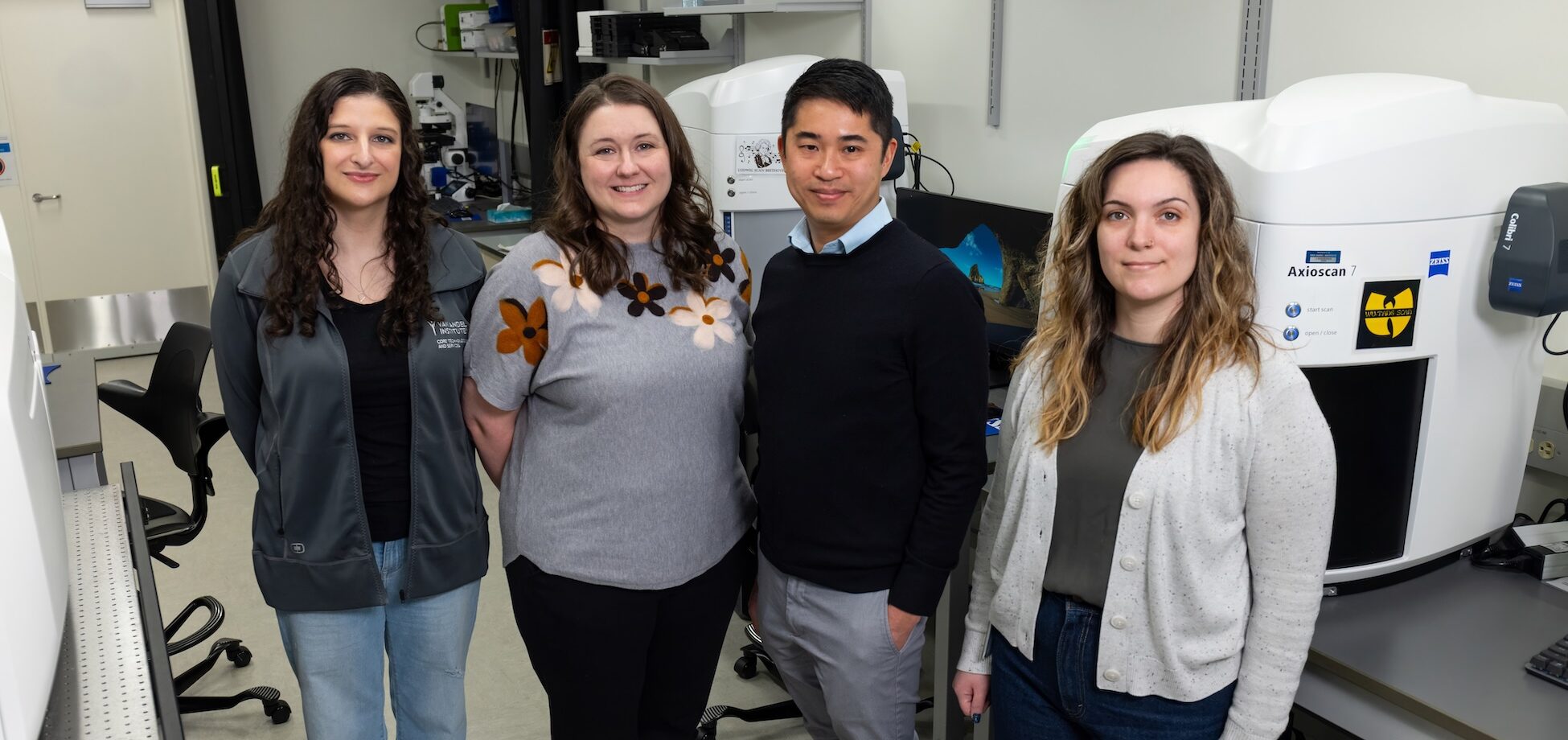

Corinne Esquibel, Ph.D.
Director, Optical Imaging Core

Lorna Cohen, Ph.D.
Core Laboratory Manager, Optical Imaging Core
Michelle Minard, B.S.
Senior Administrative Assistant II, Core Technologies and Services

Jian Wei Tay, B.Sc. (Hons I), Ph.D.
Core Bioinformatics Scientist II, Optical Imaging Core